
AI imaging second read
Every scan, read in the light of the whole patient.
PhotonMagnet reads X-ray, CT, MRI, PET and ultrasound images together with the patient's history, labs and prior reports, then gives your clinicians a full radiology report - history, technique, comparison, findings, impression and recommendations - with flagged regions, and your patients a summary they can understand.
Free demonstration with a synthetic sample patient. Not for real patient data.
Clinician
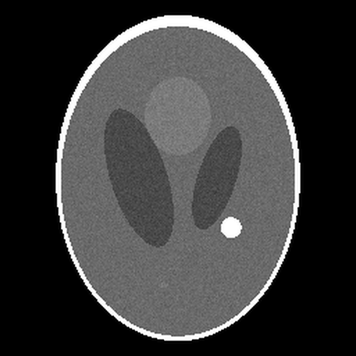
1 · Focal hyperdense lesion, left posterior parietal, ~1.4 cm, sharp margin, no mass effect.
Differential: calcified granuloma (medium) · small haemorrhage (low)
History correlation
Childhood TB exposure in the records supports a calcified granuloma.
Patient
"The scan shows one small bright spot. Spots like this are often harmless calcium…"
From upload to second read in minutes
The AI sees the images and the records at the same time, the way a radiologist who has read the chart would.
1 · Upload the study
Drop a DICOM folder, multi-frame file, JPEGs, PNGs or PDFs. DICOM is opened in your browser and patient identifiers are stripped before anything is sent.
2 · Add the history
Notes, labs, medications, prior reports - typed, PDF or photographed. Records can be added at any time and are tied to the patient and, if you like, to one study.
3 · Read the analysis
Findings tied to the exact slices and regions, a differential, correlation with the history, outlook, next steps - and a plain-language view for the patient.
For clinicians
A radiology report, ready to review
- ✓ The standard sections: clinical history, examination and technique, comparison, findings, impression, recommendations
- ✓ Findings region by region, normal structures included, compared with the patient's earlier studies
- ✓ Key findings linked to the slice and the box where they are
- ✓ Measurements to verify, never invented ones
- ✓ Differential with what argues for and against each condition
- ✓ Correlation with the history: what the records support or contradict
- ✓ Prognostic considerations
- ✓ Urgent findings flagged on the dashboard
For patients
A summary people understand
- ✓ What the scan shows, in everyday words
- ✓ What it may mean, honestly and calmly
- ✓ What usually happens next
- ✓ Questions to bring to the next appointment
- ✓ A printable page you can hand over
Built for teams
Organizations, not loose accounts
Your organization signs up once. Its administrator invites clinicians and viewers, changes their roles and disables them when they leave.
An image library
Every series, catalogued by patient, modality, body part and date, with thumbnails and a viewer with slice scrolling, window and contrast.
Walls between organizations
Every patient, image and record is reached only through its organization. Every view, upload and analysis is in the audit log.
Learn more →
Try it on the sample patient
Every new organization starts with a synthetic CT phantom and an invented history, ready to analyse.